Stroke
Stroke is a pathology associated with rapid loss of brain function due to a sudden disruption of blood supply to the brain, resulting from ischemia or haemorrhage. An ischemic stroke is caused by a blocked blood vessel and a haemorrhagic stroke is triggered by internal bleeding, for example the rupture of a cerebral aneurysm. According to the Stroke Association, 85% of strokes are caused by ischemia and the remainder are due to haemorrhage.
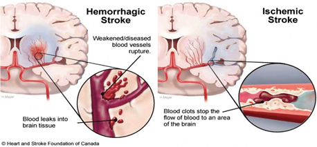
This image is taken from the Internet and the copyright belongs to the Heart and Stroke Foundation of Canada.
Recent progress in acute stroke treatment has shown that thrombolysis - the treatment of breaking up and dissolving the blood clots - increases the survival rate and reduces the disability of ischemic stroke patients. Currently, the recombinant tissue plasminogen activator (rtPA) is the only Food and Drug Administration (FDA) approved thrombolytic treatment. The rtPA has a therapeutic window of ~6 hours after the onset of ischemia.
However, the drug has some deadly side-effect such as intracranial haemorrhage. The incident rate of intracranial haemorrhage caused by the treatment ranges from 5 to 10% and the mortality rates as a result of the complication vary from 50 to 83%, about a 10-fold increase compared to the untreated group in some studies. Due to the restrictive therapeutic window and complication of rtPA administration, only approximately 5% of stroke patients are estimated to receive this therapy, though it could have potentially benefited 14 % of them.

The right movie shows the complication during rtPA treatment: a secondary hematoma can be seen shrinking as blood disgorges into the ventricles [2]; with permission from S. Karger AG, Basel.
Amide proton transfer (APT) imaging
APT imaging is a form of chemical exchange saturation transfer (CEST) that allows the detection of previously undetectable amide protons due to its low concentration, through a chemical exchange reaction with the water protons. This new and novel imaging technique has the ability to measure cellular information such as pH. Since pH drop happens prior to cerebal infarction (cell death), APT imaging has the potential to better stratify patients for rtPA to increase the benefit and to outweigh the risk of the thrombolytic treatment.
I am working closely with clinical colleagues at Oxford Acute Vascular Imaging Centre (AVIC), John Radcliffe (JR) Hospital, UK and imaging colleagues at the Functional MRI of the Brain (FMRIB). Developing the best quantification method of APT effect and facilitating APT imaging for clinical stroke imaging are the key components of my work.
Selected publications:
-
G.W.J., Harston, Y.K. Tee, N.P. Blockley et al., "Identifying the ischaemic penumbra using pH-weighted magnetic resonance imaging", Brain, 138: 36–42, 2015. [Link].
-
Y.K. Tee, G.WJ. Harston, N.P. Blockley et al., “Comparing Different Analysis Methods for Quantifying the MRI Amide Proton Transfer (APT) Effect in Hyperacute Stroke Patients”, NMR in Biomedicine, 27: 1019–1029, 2014. [Link]
-
G.W.J. Harston, Y.K. Tee et al, “Ventricular extension of intracerebral hemorrhage during intravenous thrombolysis”, Cerebrovascular Diseases, 36:324-325, 2013. [Link]
-
M.A. Chappell, M.J. Donahua, Y.K. Tee, A.A Khrapichev, N.R. Sibson, P. Jezzard and S.J. Payne, “Quantitative Bayesian Model-Based Analysis of Amide Proton Transfer MRI”, Magnetic Resonance in Medicine, 70: 556–567, 2013. [Link]